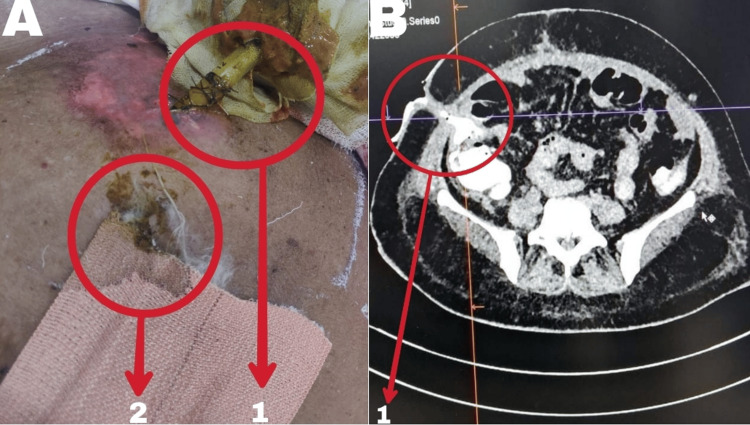
Figure 1. A) Fecal discharge from drain site and suture line on presentation; B) Contrast-enhanced computed tomography abdomen plus pelvis (CECT A+P) on presentation.
A1: fecal discharge from abdominal drain site with skin excoriation around drain site; A2: Fecal discharge from suture line with soaked dressing
B: CECT (A+P) showing complete anastomotic dehiscence and contrast extravasation from ileal loops to drain site, suggestive of enterocutaneous fistula (5.5 mm thick, 35 mm long). Multiple intra-abdominal collections, indicative of peritonitis secondary to anastomotic leak
B1: Enterocutaneous fistula (5.5 mm thick, 35 mm long)

