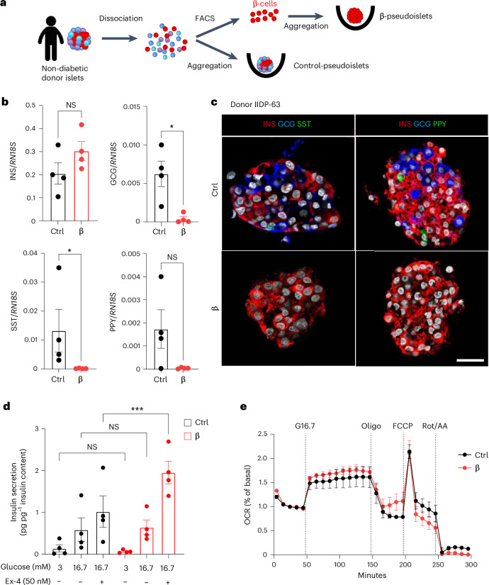
Fig. 7. Human monotypic β-pseudoislets maintain glucose-stimulated insulin secretion and islet respirometry.
a, Generation of human pseudoislets. Islets from the same donor were dissociated and sorted by FACS to obtain highly pure β-cells that were aggregated into pseudoislets. Control pseudoislets comprising all cell types were dissociated and aggregated directly. b, qPCR of INS, GCG, SST and PPY on control and β-pseudoislets. Data are normalized to housekeeping genes (RN18S); n = 4 donors. P values (Ctrl vs β): INS, P = 0.200; GCG, *P = 0.0286; SST, *P = 0.0286; PPY, P = 0.1714. NS, not significant. c, Immunofluorescence on control pseudoislets (Ctrl) or monotypic β-pseudoislets (β). INS, red; GCG, blue; SST, green (left); INS, red; GCG, blue; PPY, green (right). Scale bar, 20 µm. d, In vitro insulin secretion normalized by total insulin content in Ctrl and monotypic β-pseudoislets assessed at basal (3 mM, low glucose (LG)) or stimulatory (16.7 mM, high glucose (HG)) glucose concentrations in the presence or absence of the GLP-1 agonist Ex-4. n = 4 donors. P values (Ctrl vs β): LG, P = 0.1831; HG, P > 0.999; HG + Ex-4, ***P = 0.0002. NS, not significant. e, Islet respirometry by Seahorse XF96 bioanalyser. OCR traces of human Ctrl or β-only pseudoislets. Glucose (16.7 mM; G16.7), oligomycin (2.5 µM; Oligo), FCCP (2 µM) and Rot/AA (3 µM) were injected at the indicated time points. Basal measurement was performed at 3 mM glucose before injections. OCR was normalized by insulin content of each well and represented as % of basal. n = 3 independent donors. Two to five wells per condition per donor, ten pseudoislets per well; Two-way ANOVA with Sidak’s multiple comparisons test. Both male and female human donors were used. All data are shown as mean ± s.e.m. Unless otherwise indicated, P values are from two-tailed Mann–Whitney tests.

