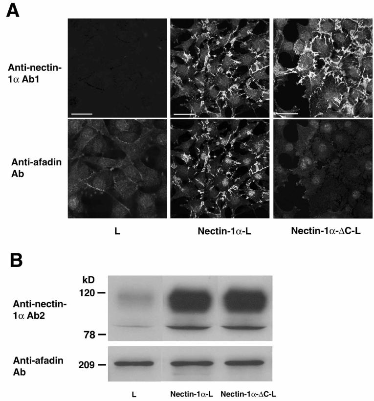
FIG. 1.
Interaction of afadin with nectin-1α, but not with nectin-1α-ΔC. (A) Immunofluorescence microscopy. L, nectin-1α-L, and nectin-1α-ΔC-L cells were doubly stained with the polyclonal anti-nectin-1α Ab1 and the monoclonal anti-afadin Ab. There was nuclear staining with this monoclonal anti-afadin Ab, but its significance is not clear. Bars, 10 μm. (B) Western blotting. Each cell lysate of L, nectin-1α-L, and nectin-1α-ΔC-L cells (20 μg of protein each) was subjected to SDS-PAGE (10% polyacrylamide gel), followed by Western blotting with the polyclonal anti-nectin-1α Ab2 and the monoclonal anti-afadin Ab. These results are representatives of three independent experiments.

