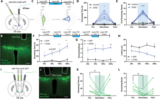
Figure 2.

ZITH neurons bidirectionally regulate self‐grooming. A) Experimental diagram of optogenetic activation of ZITH neurons; B) a post‐hoc image illustrating the expression of AAVs and the location of optical fiber (scale bar, 200 µm); C) schematic diagram of optogenetic stimulation strategy, and summary radar plot of behaviors during laser exposure (control: N = 6 mice, ChR2: N = 7 mice); D,E) total grooming time and the number of grooming bouts in 5 min before, during, and after optogenetic stimulation of ZITH neurons (control, N = 3 mice, and ChR2: N = 4 mice, p = 0.032 and p = 0.0002, repeated measurements two‐way ANOVA). F–H) experimental strategy, a total grooming time, the number of grooming bouts, and latencies in the 90s induced by varying stimulation frequencies (N = 3 from control, and N = 4 from ChR2 mice). I) experimental diagram of optogenetic inhibition of ZITH neurons; J) a post‐hoc image illustrating the expression of AAVs and the location of optical fiber (scale bar, 200 µm); K,L) total grooming time and the number of grooming bouts in 5 min before, during, and after optogenetic inhibition of ZITH neurons (N = 5 mice, p = 0.046 and p = 0.031, paired t‐test). *p < 0.05, ***p < 0.001.
