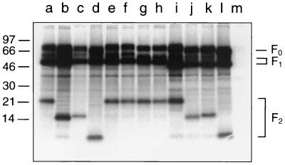
FIG. 2.
Electrophoretic mobilities of the F protein mutants. MVA-T7-infected BSR-T7/5 cells were transfected with recombinant pTM1 plasmids (lane a, parental F; lane b, N27Q; lane c, N70Q; lane d, N27/70Q; lane e, N116Q; lane f, N120Q; lane g, N116/120Q; lane h, N126Q; lane i, N500Q; lane j, N27/500Q; lane k, N70/500Q; lane l, N27/70/500Q; and lane m, pTM1). The cells were metabolically labeled with [35S]methionine-[35S]cysteine, F protein was immunoprecipitated from the cell lysates, and the immunoprecipitates were separated by Tricine–SDS–10% polyacrylamide gel electrophoresis under reducing conditions. The relative positions of standard proteins of the indicated molecular masses (in kilodaltons) are shown on the left.

