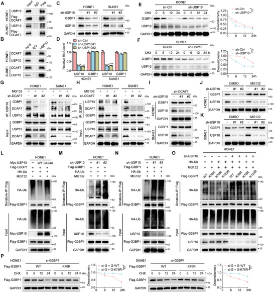
Figure 4.

DCAF7 recruits USP10 to deubiquitylate and stabilize G3BP1. A,B) IP (with an anti‐FLAG antibody or IgG) was conducted to validate the interaction between DCAF7 and USP10 in SUNE1 and HONE1 cells transfected with Flag‐DCAF7. C,D) Protein and mRNA levels of G3BP1 in HONE1 and SUNE1 cells with or without USP10 knockdown. Mean (n = 3) ± s.d. One‐way ANOVA, *** p < 0.001. E,F) Protein level of G3BP1 in HONE1 and SUNE1 cells with or without USP10 knockdown following CHX treatment (100 µg mL−1) for the indicated times. Mean (n = 3) ± s.d. Two‐way ANOVA, ** p < 0.01. G,H) IP (with an anti‐USP10 or anti‐G3BP1 antibody) and IB of G3BP1, DCAF7 and USP10 in HONE1 and SUNE1 cells transduced with sh‐control or sh‐DCAF7 following MG132 treatment (10 µm, 6 h). I) IB of G3BP1, USP10 and GAPDH in DCAF7‐knockdown HONE1 and SUNE1 cells transduced with sh‐control or sh‐USP10. J,K) IB of G3BP1, USP10 and GAPDH in HONE1 and SUNE1 cells transduced with sh‐control or sh‐USP10 following MG132 treatment (10 µm, 6 h). L–N) Denaturing IP with an anti‐Flag antibody and IB of HA‐Ub, Flag‐G3BP1, Myc‐USP10 and GAPDH in HONE1 and SUNE1 cells transfected with the indicated plasmids following MG132 treatment (10 µm, 6 h). O) Denaturing IP with an anti‐Flag antibody and IB of HA‐Ub, Flag‐G3BP1, USP10 and GAPDH in HONE1 cells transfected with the indicated plasmids following MG132 treatment (10 µm, 6 h). P) Protein level of Flag‐G3BP1 in HONE1 and SUNE1 cells transfected with indicated siRNA and plasmids following CHX treatment (100 µg mL−1) for the indicated times. Mean (n = 3) ± s.d. Two‐way ANOVA, ** p < 0.01. The unprocessed images of the blots are shown in Figure S10 (Supporting Information).
