Figure 5.

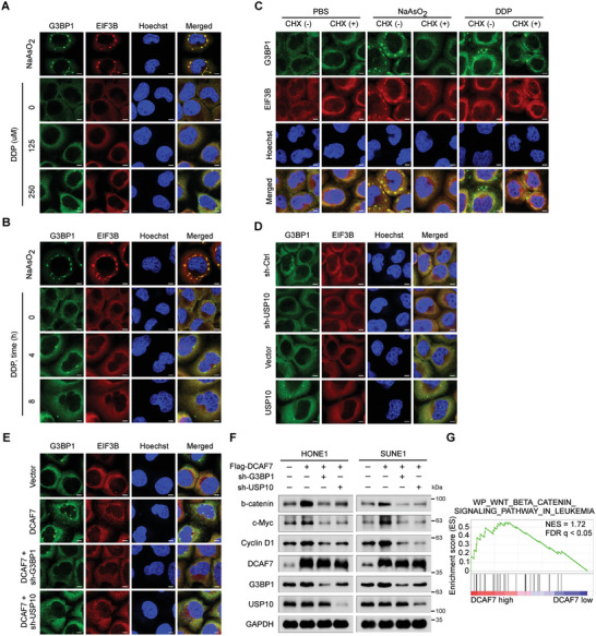
DCAF7 facilitates cisplatin‐induced formation of SG‐like structures. A,B) IF staining (with an anti‐G3BP1 or anti‐EIF3B antibody) of HONE1 cells subjected to stress induction via cisplatin (0, 125, or 250 µm) for 4 h (A) or to cisplatin treatment (250 µm) for 0, 4, or 8 h (B). As a positive control, cells were treated with 500 µm sodium arsenite (NaAsO2) for 1 h to induce robust SG formation. C) Cells were incubated with NaAsO2 (500 µm for 1 h) or cisplatin (250 µm for 4 h) and then treated with CHX (100 µg mL−1 for 30 min) for forced SG disassembly, and immunostaining for G3BP1 and EIF3B was then performed. D,E) IF staining (with an anti‐G3BP1 or anti‐EIF3B antibody) of HONE1 cells transfected with the indicated plasmids following cisplatin (250 µm) treatment for 4 h. The scale bar corresponds to 5 µm (A–E). F) IB of β‐catenin, c‐Myc, Cyclin D1, G3BP1, DCAF7, USP10 and GAPDH in HONE1 and SUNE1 cells transfected with the indicated plasmids following cisplatin treatment (10 µg mL−1) for 24 h. G) GSEA of the GSE102349 dataset demonstrated positive enrichment of genes associated with Wnt/β‐catenin signalling in response to DCAF7 overexpression. The unprocessed images of the blots are shown in Figure S10 (Supporting Information).
