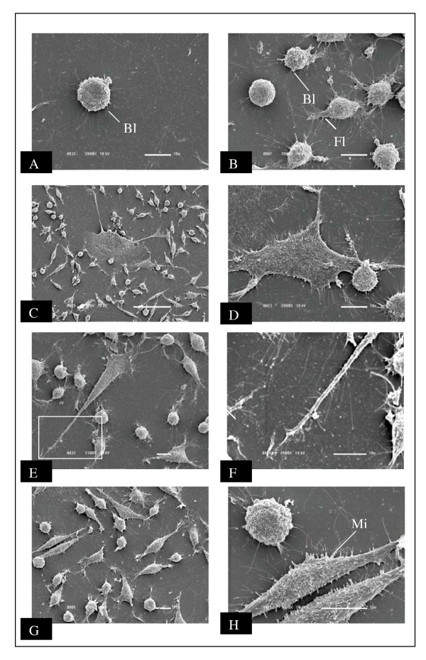
Figure 3.
Scanning electron micrograph of NG97 cell line. A, B: small rounded cells presenting blebs (Bl) and filopodia (Fi) on their surfaces; C, D, E: dendritic-like cells with extensive cytoplasmatic prolongations. The area in the rectangle is shown at higher magnification in F; G: culture with two morphologic distinct cellular types; H: fibroblastic-like cells presenting microvilli (Mi) on the membrane surface.

