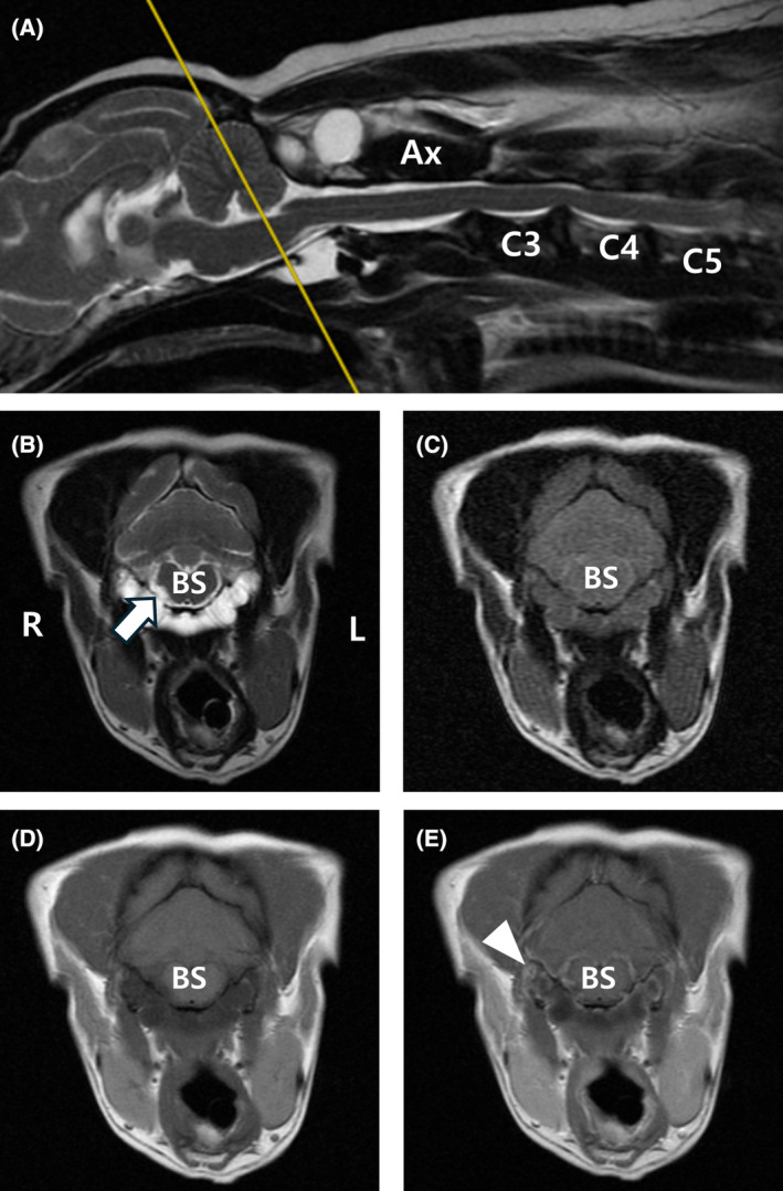
FIGURE 1.

Magnetic resonance images of cystic lesions at the level of the brainstem. Sagittal (A) and transverse (B) T2‐weighted, transverse FLAIR (C), T1‐weighted (D), and T1‐weighted post‐contrast (E) magnetic resonance images. The lesion is observed hyperintense in T2‐weighted images (A, B). In the T2‐weighted FLAIR image, hyperintensity is observed compared with CSF (C), and there is mild peripheral enhancement (white arrowhead) in T1‐weighted post‐contrast images (E). At the brainstem level, the cystic lesion was located in the ventral region of the vertebra and was observed to be bilateral, encircling the atlas and resulting in slight compression of the right‐sided hypoglossal nerve (white arrow). Ax, axis; BS, brainstem; FLAIR, fluid‐attenuated inversion recovery; L, left; R, right.
