FIGURE 3.

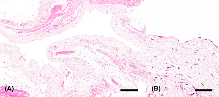
Ganglion cyst, hematoxylin and eosin. Large cystic structure with an empty lumen surrounded by an edematous fibrous connective tissue wall lined by flattened, well‐differentiated cells. Bar = 800 μm (A). Higher magnification of the flattened cells lining the cystic structure. Bar = 100 μm (B).
