
FIGURE 2.
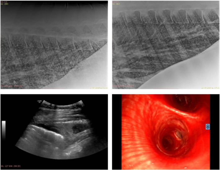
Diagnostic imaging findings from 2 horses with systemic leptospirosis. Top images: thoracic radiographs of a horse with epistaxis and pulmonary hemorrhage. There is a generalized mixed lung pattern caudodorsally with some improvement from Day 2 (left) to Day 10 (right). Bottom left: thoracic ultrasonography of the same horse as above. There is mild irregularity of the pleura and pleural effusion of mixed echogenicity. Bottom right: tracheobronchoscopy of another horse with epistaxis and pulmonary hemorrhage. There is severe hemorrhage in the trachea and main bronchi.