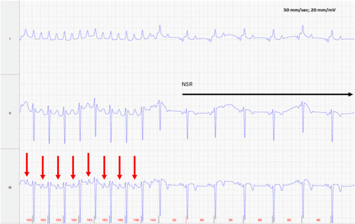
FIGURE 5.

Foal 2, Day 1, at time of conversion to normal sinus rhythm (NSR). Lead II is in a base apex configuration. Lead I is a nontraditional lead and Lead III is calculated from Leads I and II. The atrial activity (red arrows) has become more organized before conversion with an atrial rate of 160 bpm just before conversion. For all ECGs, the electrodes are placed around the girth area with the left leg electrode on the sternum, the left arm electrode on left side of the thorax slightly dorsal to level of the cardiac apex, the right arm electrode over the right thorax approximately 6 cm below the dorsal midline, and the right leg electrode over the left thorax approximately 6 cm below the dorsal midline.
