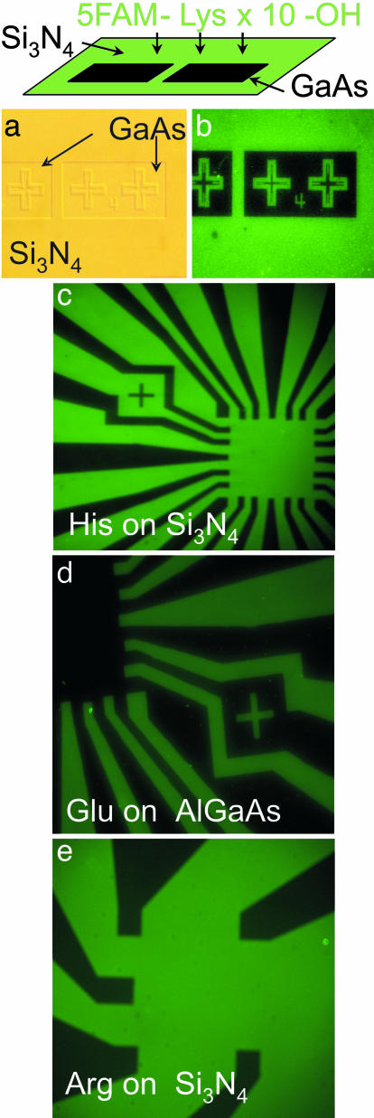
Fig. 1.
Adhesion of single amino acid constituted peptides to various inorganic surfaces as observed by using fluorescence microscopy. (a and b) Differential amino acid adhesion to inorganic surfaces as demonstrated in normal phase contrast (a) and fluorescence micrograph (b) of patterned structure used to test differential peptide adhesion. This structure is Si3N4 (deposited with plasma-enhanced chemical vapor deposition) on (100) GaAs with the pattern due to areas of the Si3N4 that have been removed by dry etching (reactive ion etching). This sample was then placed in a solution of peptide containing 10 Lys, N-terminated by a fluorescence molecule, then water washed. No evidence of fluorescence is apparent on the exposed GaAs, but the Si3N4 surface fluoresces homogeneously, corresponding to a peptide density of ≈2 × 10⁴ peptides per μm2. (c–e) Fluorescence micrographs of amino acid (10-mer peptides) adhesion to multiple surfaces. The dark elements in each micrograph are the GaAs substrates to which minimal adhesion is observed.

