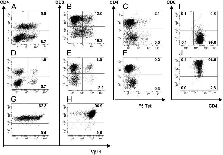
Fig. 1.
Retroviral gene transfer of TCR chains, purification of TCR-expressing CD4+ and CD8+ T cells, and retroviral gene transfer of CD8α.(A–F) Flow cytometric analysis of viable CD3+ murine splenocytes 2 days after retroviral transfer with F5 TCRα and TCRβ genes (A–C) or mock transduction (D–F). Cells were stained with PE-anti-Vβ11, PE-labeled NP tetramer, FITC-anti-CD4, or allophycocyanin-anti-CD8. (G and H) Flow cytometric analysis of purified CD4+ and CD8+ T cells. Forty-eight hours after transduction, TCR-td bulk T cells were sorted into CD4+ and CD8+ T cells by using magnetic beads, followed by enrichment for Vβ11+ cells 1 day later. Cells were stained with PE-anti-Vβ11, FITC-anti-CD4, and/or allophycocyanin-anti-CD8, and quadrants were set based on anti-CD4 and anti-CD8 stained mock-td cells. (I and J) Flow cytometric analysis of purified TCR-td CD4+ T cells (I) and TCR-td CD4+8+ T cells (J) after retroviral cotransfer of F5 TCRα, TCRβ, and murine CD8α genes. After cotransfer of CD8α, >95% of CD4+ T cells coexpressed CD8α (TCR-td CD4+8+ T cells).

