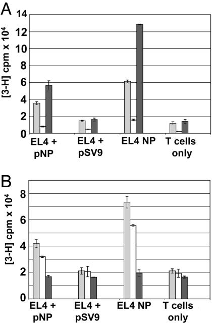
Fig. 3.
In vitro proliferative potential and IL-2 production of TCR-td CD4+ T cells and TCR-td CD4+8+ T cells. (A) TCR-td CD4+8+ T cells proliferated poorly in response to peptide-presenting tumor cells (white bars) compared with TCR-td CD4+ T cells (gray bars) and TCR-td CD8+ T cells (black bars). The proliferative potential of the TCR-td T cell populations correlated well with IL-2 production. (B) TCR-td CD4+8+ T cells (white bars) secreted reduced levels of IL-2 in response to EL4 NP tumor cells as compared with TCR-td CD4+ T cells (gray bars). TCR-td CD8+ T cells (black bars) did not secrete IL-2 in response to any of the targets. Proliferation of the IL-2-dependent cell line CTLL was used to measure the IL-2 content in the supernatant taken from the stimulated T cells.

