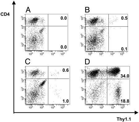
Fig. 5.
Detection and in vivo persistence of TCR-td T cells in tumor-bearing mice. Shown is the analysis of splenocytes prepared from mice in Fig. 4A at the time they were killed because of tumor burden. Adoptively transferred T cells were identified by triple staining with antibodies specific for Thy1.1, Vβ11, and CD4. Representative examples are shown for each group of mice, and all FACS plots display viable lymphocytes gated on Vβ11+ T cells. (A) No Thy1.1+ Vβ11+ T cells were identified in four tumor-bearing PBS-treated control mice. (B) In four mice treated with mock-td CD4+ T cells, between 0.5% and 1.7% of the gated Vβ11+ cells were the CD4+ Thy1.1+ transferred T cells. (C) Similar numbers of TCR-td CD4+8+ cells were detected in three mice (0.5–3.3%) suggesting a lack of antigen-driven expansion of the TCR-td CD4+8+ as compared with the Mock-td CD4+ T cells. (D) Analysis of the spleen of a tumor-bearing animal that received TCR-td CD4+ T cells showed a large expansion of transferred Thy1.1+ CD4+ T cells (34.0% of gated Vβ11+), together with an expansion of the cotransferred Thy1.1+ CD8+ T cells (13.6% of gated Vβ11+). Staining of lymph node cells of mice in the four treatment groups showed similar levels of transferred Thy1.1+ Vβ11+ CD4+ T cells (data not shown).

