Fig. 2.
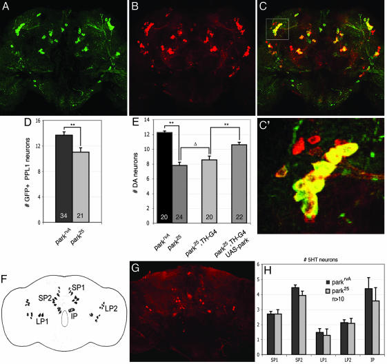
Neuron loss in parkin mutants is specific to DA neurons and results primarily from loss of a cell-autonomous requirement of parkin.(A–C) Expression of GFP driven by TH-GAL4 (A) and anti-TH (B) staining reveals a high degree of colocalization of signals (C). The boxed area in C marks the PPL1 cluster and is magnified in C′.(D) Analysis of GFP-positive neurons in 20-day-old adult parkin mutants and age-matched WT controls bearing the TH-GAL4 driver and a UAS-GFP transgene confirms the cell loss in the PPL1 DA neuron cluster of parkin mutants (**, P < 0.005, Student's t test). (E) DA neuron loss is significantly reduced by expression of a parkin transgene using the TH-GAL4 driver compared with parkin mutant or parkin mutants bearing the TH-GAL4 driver alone. Expression of GAL4 from the TH-GAL4 driver did not significantly affect DA neuron viability. (**, P < 0.001; Δ, P = 0.3) (F) Diagram shows all major serotonergic neurons in the adult brain. SP, subaesophageal; LP, lateral protocerebral; IP, inferior medial protocerebral. (G) Representative confocal micrograph of a WT adult brain stained with anti-5-hydroxytyrosine to reveal serotonergic neurons. (H) No significant difference in the number of serotonergic neurons is detectable in 20-day-old adult parkin mutants relative to age-matched controls, indicating that neuron loss is specific to a subset of DA neurons. Statistical significance was calculated by using ANOVA and Bonferroni's post hoc test for planned comparisons. Numbers shown in histograms refer to the number of brains used for neuron counts. park25 or parkrvA homozygotes were used in all analyses of neuronal viability.

