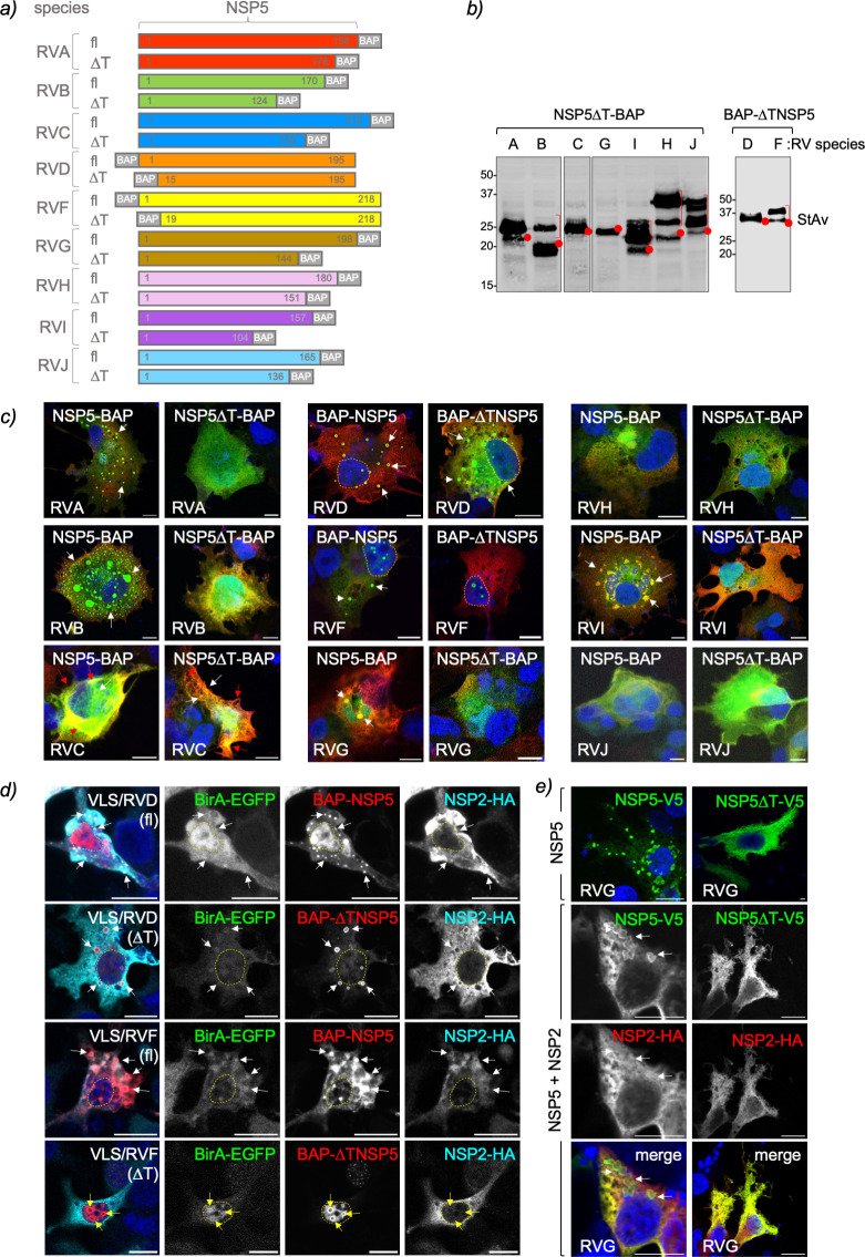
Fig 3.
NSP5 ordered region, tail, is required for VLS formation among RV species. (a) Schematic representation of RV species A to J of full-length (fl) NSP5 and NSP5 with deleted tail region (∆T) fused to a BAP tag at the N- or C-terminus as indicated. b) Immunoblotting of MA/cytBirA cells expressing NSP5∆T-BAP (RVA to RVC and RVG to RVJ) and BAP-∆TNSP5 (RVD and RVF). The membrane was incubated with streptavidin-IRDye800. The red dots indicate the predicted molecular weight of the proteins. The red bracket shows a slow migration pattern of the protein. (c) Merged immunofluorescence images of MA/cytBirA cells co-expressing NSP2-HA with NSP5 or NSP5∆T fused to BAP tag. At 16 hpt, the cells were fixed and immunostained for the detection of NSP5 or NSP5∆T fused to BAP tag (StAv, green) or NSP2-HA (anti-HA, red). Nuclei were stained with DAPI (blue). The scale bar is 10 µm. (d) Immunofluorescence images of LMH cells co-expressing BirA-EGFP and NSP2-HA with BAP-NSP5 or BAP-∆TNSP5 of RVD and RVF. The cells were fixed at 16 hpt and immunostained for detection of BirA-EGFP (green, second column), NSP5 (StAv, red, third column), and NSP2 (anti-HA, cyan, fourth column). Nuclei were stained with DAPI (blue). The scale bar is 10 µm. A merged image is presented in the first column. (e) Immunofluorescence of LMH cells expressing NSP5-V5/G fl (NSP5-V5) or ∆T (NSP5∆T-V5) alone or together with NSP2-HA/G. After fixation, the cells were immunostained for the detection of NSP5 (anti-V5, green) and NSP2 (anti-HA, red). Nuclei were stained with DAPI (blue). The scale bar is 10 µm. A merged image for the co-expression of NSP5-V5 with NSP2-HA is shown. In (d) and (e), the white and red arrows point to globular and filamentous VLSs, respectively. The yellow arrows point to nuclear inclusions, and discontinued yellow lines label the nucleus position.

