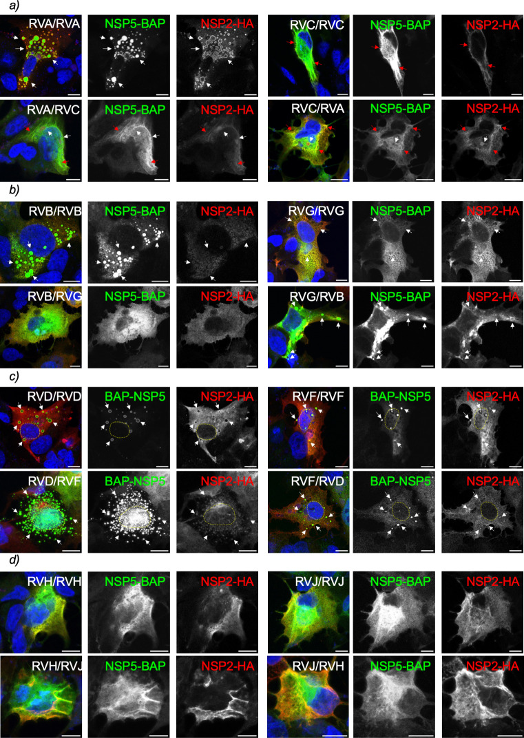
Fig 7.
Heterologous formation of VLS among closely related RV species. Immunofluorescence images of MA/cytBirA cells co-expressing NSP5 tagged to BAP and NSP2-HA of closely related RV species A and C (a), B and G (b), D and F (c), and H and J (d). After fixation, the cells were immunostained for the detection of NSP5 (StAv, green) and NSP2 (anti-HA, red). The nuclei were stained with DAPI (blue). The indication at the top right corner corresponds to the RV species of NSP5 and NSP2, respectively. The scale bar is 10 µm. The white and red arrows point to globular and filamentous VLSs, respectively. The yellow lines label the nucleus position.

