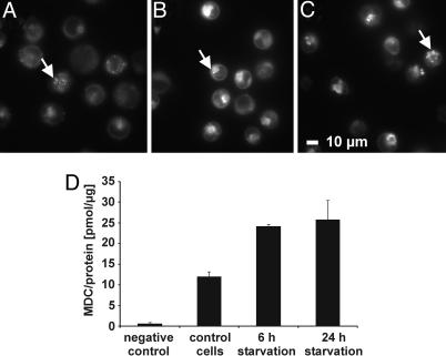
Fig. 1.
Starvation enhances the level of autophagic vacuoles. Autophagic vacuoles were stained with the specific dye MDC (13) and analyzed by fluorescence microscopy or fluorescence spectroscopy. Awells cells were incubated for 24 h in DMEM (control cells) (A), 6 h HBSS (B), or 24 h HBSS (starved cells) (C), subsequently, for 10 min with MDC, washed, and immediately analyzed by fluorescence microscopy. Autophagic vacuoles are marked with an arrow. (D) Intracellular MDC measurement by fluorescence spectroscopy; unstained cells were used as negative control.

