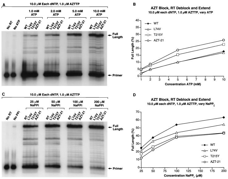
FIG. 4.
(A) The PBS primer was 5′ end labeled and annealed to the template as described in Materials and Methods. The 3′ end of the primer was blocked by the addition of an AZT residue. The ability of wild-type HIV-1 RT and the RT variants to remove the blocking AZT residue (deblocking) and to extend the freed end of the primer was tested in the presence of 10.0 μM concentrations of each dNTP, 1.0 μM AZTTP, and varying concentrations of ATP (1.0, 2.0, 5.0, and 10.0 mM) as the pyrophosphate donor. In the cell, nucleoside analogs will be present in the triphosphate form, and after a primer is deblocked there is a possibility that HIV-1 RT will add another nucleoside analog back on to the 3′ end of the primer rather than the normal dNTP, which in this case is dTTP. The addition of AZTTP to the reaction mixture is meant to reflect what can occur within the cell. A control lane with no added wild-type RT shows the pattern of the starting template-primer. A control lane to which has been added HIV-1 RT but no ATP indicates the amount of extendable primer. This could result from primer which did not get blocked by an AZT residue or from a low level of deblocking by the enzyme using the dNTPs as the pyrophosphate donor or a combination of both processes. The locations of the starting PBS primer and the fully extended primer are marked. (B) The gel in panel 4A was scanned by a PhosphorImager. In each lane, the amount of radioactivity in the full-length product was divided by the total amount of radioactivity to determine the percentage of full-length product. This value was plotted versus the level of ATP present in the reaction mixture. The percentage of full-length product in the No ATP control lane indicates that the background level is very low (<1.0%). (C) The 3′ end of the primer was blocked by the addition of an AZT residue, and the ability of wild-type HIV-1 RT and the RT variants to remove the blocking AZT residue and extend the freed end of the primer was tested in the presence of 10.0 μM concentrations of each dNTP, 1.0 μM AZTTP, and varying concentrations of NaPPi (25.0, 50.0, 100.0, and 200.0 μM) as the pyrophosphate donor. The locations of the starting PBS primer and the fully extended primer are marked. (D) The gel in panel C was scanned by a PhosphorImager. In each lane, the amount of radioactivity in the full-length product was divided by the total amount of radioactivity to determine the percentage of full-length product. This value was plotted versus the level of NaPPi present in the reaction mixture. The percentage of full-length product in the no NaPPi control lane indicates that the background level is very low compared to that for the reactions where NaPPi is present.

