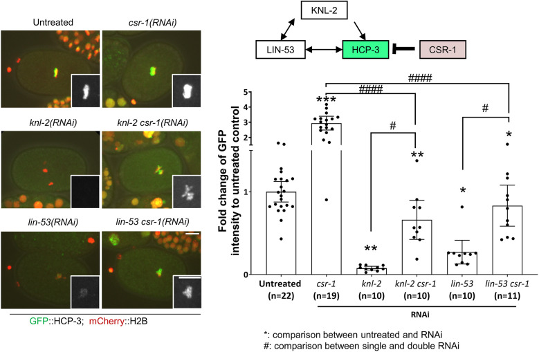
Fig. 5.
Depletion of CSR-1 promotes HCP-3 chromatin localization in embryos depleted of HCP-3 loading factors. Representative images and quantification of the mean GFP::HCP-3 intensity of the metaphase plate in one-cell embryos with RNAi treatments for depletion of CSR-1, KNL-2 and LIN-53 alone or in combination, as indicated. Inset images show magnified views of the GFP::HCP-3 signal on the metaphase plate. Diagram shows the localization dependency relationship among the proteins. Scale bars: 10 μm; insets, 5 μm. Data are presented as mean±95% confidence interval. The n values shown represent the number of one-cell embryos analyzed. *P<0.05; #P<0.05; **P<0.01; ***P<0.001; ####P<0.0001 (two-tailed unpaired t-test). The representative images of untreated control, knl-2 csr-1(RNAi) and lin-53 csr-1(RNAi) are also shown in Fig. S6A for comparison with PRG-1-depleted embryos as part of the same experiment.

