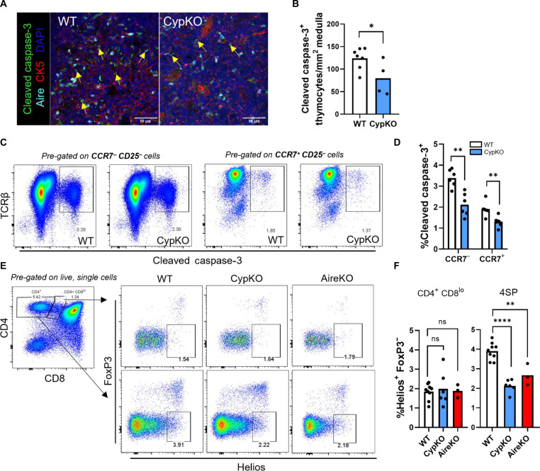
Fig. 3. Decreased thymocyte apoptosis in CypKO mice.
(A and B) IF microscopy images of cleaved caspase-3 staining in the thymic medulla in small CK5− cells (A) and quantification of positive cell density (B) in WT versus CypKO from the entire thymic cross sections. (C and D) Representative flow cytometry plots of cleaved caspase-3+ cells in WT versus CypKO CCR7− CD25− (left) or CCR7+ CD25− thymocytes (right) (C) and quantification of cleaved caspase-3+ thymocytes (D). (E and F) Representative flow cytometry plots of Helios+ FoxP3− CD4+ CD8lo (top) or 4SP (bottom) thymocytes in WT versus CypKO or Aire KO thymi (E) and summary data (F). All experiments were performed with age-matched (male and female) 8- to 12-week-old mice. Each dot represents an individual mouse. Statistics: Unpaired parametric t tests. *P < 0.05; **P < 0.01; ****P < 0.0001.

