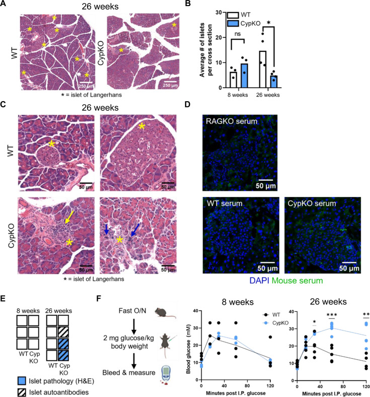
Fig. 4. Evidence of disrupted pancreatic function and islet pathology in aged CypKO mice.
(A) Representative H&E images of pancreases from 26-week-old WT and CypKO mice. Islets are marked with an asterisk. (B) Quantification of the average number of islets from duplicate whole-tissue cross section scans from 8- and 26-week-old WT and CypKO mice. (C) High-magnification images of WT or CypKO islets from duplicate mice. Immune cell infiltration is indicated with a yellow arrow, and pycnotic nuclei are indicated with blue arrows. (D) Representative images of autoantibody staining of islets with 26-week-old CypKO, WT, or Rag1KO serum samples. (E) Summary of indicated phenotypes in each mouse. Empty boxes indicate that no pathology or autoantibody staining was observed. (F) Experimental design for the glucose tolerance test (left) and blood glucose levels in 8- or 26-week-old WT versus CypKO mice after intraperitoneal (I.P.) glucose administration. All experiments were performed with age-matched (male or female) 8- or 26-week-old mice, except for histology for aged animals, which was performed on male mice exclusively. O/N, overnight. Statistics: Each dot represents an individual mouse. Unpaired parametric t tests. *P < 0.05; **P < 0.01; ***P < 0.001.

