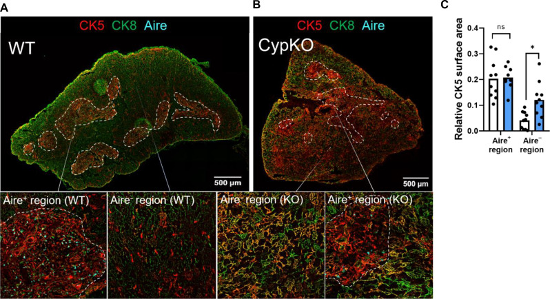
Fig. 5. Disorganized thymic architecture in CypKO mice.
(A) Representative images of a WT thymic cross section with Aire staining restricted to areas of concentrated CK5 staining (medulla). (B) Representative images of a CypKO thymic cross section showing disseminated CK5 staining in Aire− regions. (C) Quantification of CK5 staining in the medulla (defined as Aire+ regions) and cortex (defined as Aire− regions). All experiments were performed with age-matched (male and female) 8- to 12-week-old mice. Each dot represents an individual mouse. Statistics: Unpaired parametric t tests. *P < 0.05.

