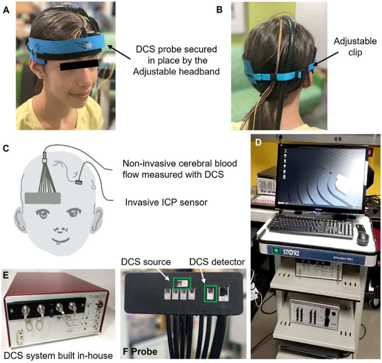
FIG. 1.

Details of DCS instrumentation and measurement setup. A–B: Probe secured on the left forehead with the adjustable headband. C: Probe placement on the right forehead of a patient, where the ICP monitor is surgically inserted in the left frontal lobe. D: Hospital cart with instruments placed. E: Custom-made DCS device. F: Custom-made DCS optical probe. The green regions refer to the DCS source and detector.
