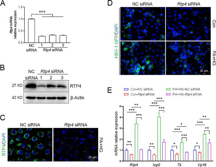
Figure 5.
Rtp4 knockdown restricted HSV-1 infection in a T2D cell model. Knockdown of Rtp4 by specific siRNAs was detected by quantitative PCR and Western blot in HCECs of the control group (A, B) and immunofluorescence staining in HCECs of the T2D group (C). (D) After 24 hours of HSV-1 infection, representative images showing GFP-labeled HSV-1 infection with NC siRNA and Rtp4 siRNA. MOI = 1. (E) Quantitative PCR analysis of HSV-1 viral genes and Rtp4 gene in Figure 5C. The h-β-actin was used as an internal control. NC, negative control. *P < 0.05, **P < 0.01, ***P < 0.001.

