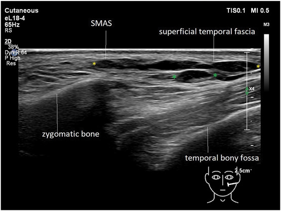
Figure 6.

Ultrasound (grayscale, oblique view) of the temple area depicting filler material injected into the SMAS distributed into the superficial temporal fascia (yellow markers) and some filler material in the deep interfascial plane (green markers [Philips affinity 70 18 MHz]).
