. 2024 Sep 25;13:RP98238. doi: 10.7554/eLife.98238

Figure 1. ieCtnnb1 is an intestinal enhancer of Ctnnb1.

(A) Schematic representation of the upstream region of mouse Ctnnb1 gene and the location of ieCtnnb1 (6,399 bp, pink shading), which is marked by H3K27ac and H3K4me1 peaks, and DNase I hypersensitivity in small intestine and large intestine of 8-week-old mice. The sequence conservation of the indicated species is shown at the bottom as vertical lines. Data were obtained from ENCODE. Locations of single-guide RNAs (sgRNAs) for generating ieCtnnb1 knockout mice were marked. (B) Top left: a schematic illustration showing that the knock-in reporter construct carries the Shh promoter, ieCtnnb1 core region sequences (2,153 bp), and the LacZ reporter gene. Top right: X-Gal staining (blue) of the gastrointestinal (GI) tract of an 8-week-old H11i.enh mouse. Bottom: X-Gal staining (blue) of the small intestine (left) and colon (right) of an 8-week-old H11i.enh mouse. Boxed areas were enlarged at top-right corners. (C) Representative images of small intestinal crypts co-labeled by X-Gal with OLFM4 (left), and X-Gal with Lysozyme (right). (D–E) Representative images of whole body (d) and GIs (e) of 8-week-old male wildtype (WT) and Ctnnb1Δi.enh mice. (F) Comparison of the body weight of 8-week-old male WT (n=13) and Ctnnb1Δi.enh (n=13) mice. (G–H) Measurements of small (G) and large (H) intestine length of 8-week-old male WT (n=6) and Ctnnb1Δi.enh (n=6) mice. (I–J) Relative mRNA levels of Ctnnb1 in small (I) and large (J) intestinal crypts of WT (n=6) and Ctnnb1Δi.enh (n=6) mice. (K–L) Left: immunoblotting of nuclear (K) and cytoplasmic (L) β-catenin, GAPDH, and H3 of small intestinal crypts of WT (n=3) and Ctnnb1Δi.enh (n=3) mice. Right: histograms showing protein levels of β-catenin normalized to H3 (K) or GAPDH (L) levels. Values of WT were set as ‘1’. (M) Heatmap of indicated Wnt target genes and gene set enrichment analysis (GSEA) of Wnt signaling pathway according to transcriptome profiles of small intestinal crypts of WT (n=3) and Ctnnb1Δi.enh (n=3) mice. (N) Quantitative reverse transcription PCR (RT-qPCR) showing relative mRNA levels of indicated Wnt target genes (Axin2, Lgr5, and Mmp7) in small intestinal crypts of WT (n=3) and Ctnnb1Δi.enh (n=3) mice. Scale bars, 1 cm (B, top; D and E), 100 μm (B, bottom), 10 μm (B, magnified views; C). Quantification data are shown as means ± SEM, statistical significance was determined using an unpaired two-tailed Student’s t-test (F–L). Quantification data are shown as means ± SD, statistical significance was determined using Multiple t-tests – one per row (N). *p<0.05, **p<0.01, ***p<0.001, and ****p<0.0001. ns, not significant. NES: normalized enrichment score.

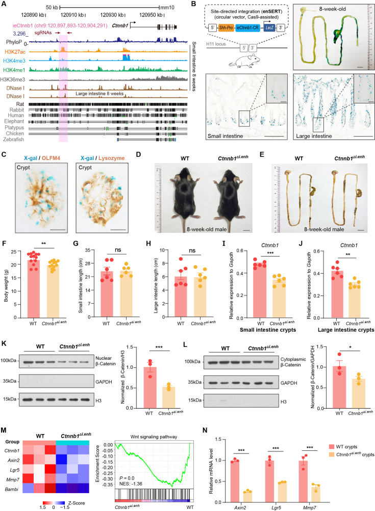
Figure 1—source data 1. Numerical data for Figure 1F.
Figure 1—source data 2. Numerical data for Figure 1G.
Figure 1—source data 3. Numerical data for Figure 1H.
Figure 1—source data 4. Numerical data for Figure 1I.
Figure 1—source data 5. Numerical data for Figure 1J.
Figure 1—source data 6. Numerical data for Figure 1K.
Figure 1—source data 7. Numerical data for Figure 1L.
Figure 1—source data 8. Numerical data for Figure 1N.
Figure 1—source data 9. Uncropped and labeled gels for Figure 1.
Figure 1—source data 10. Raw unedited gels for Figure 1.

Figure 1.

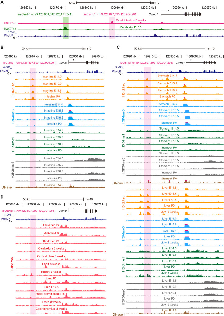
Figure 1—figure supplement 1. ieCtnnb1 is a putative intestinal enhancer upstream of Ctnnb1.

Figure 1—figure supplement 1.

(A) Schematic representation of the upstream region of mouse Ctnnb1 gene and locations of enhancer neCtnnb1 (green shading) and putative enhancer ieCtnnb1 (pink shading). Data were obtained from ENCODE. (B–C) Enrichment of indicated signals in developing intestine (B), stomach, and liver (C) were shown. Data were obtained from ENCODE. (D) Enrichment of H3K27ac chromatin immunoprecipitation sequencing (ChIP-seq) signals in indicated mouse tissues. Data were obtained from ENCODE.
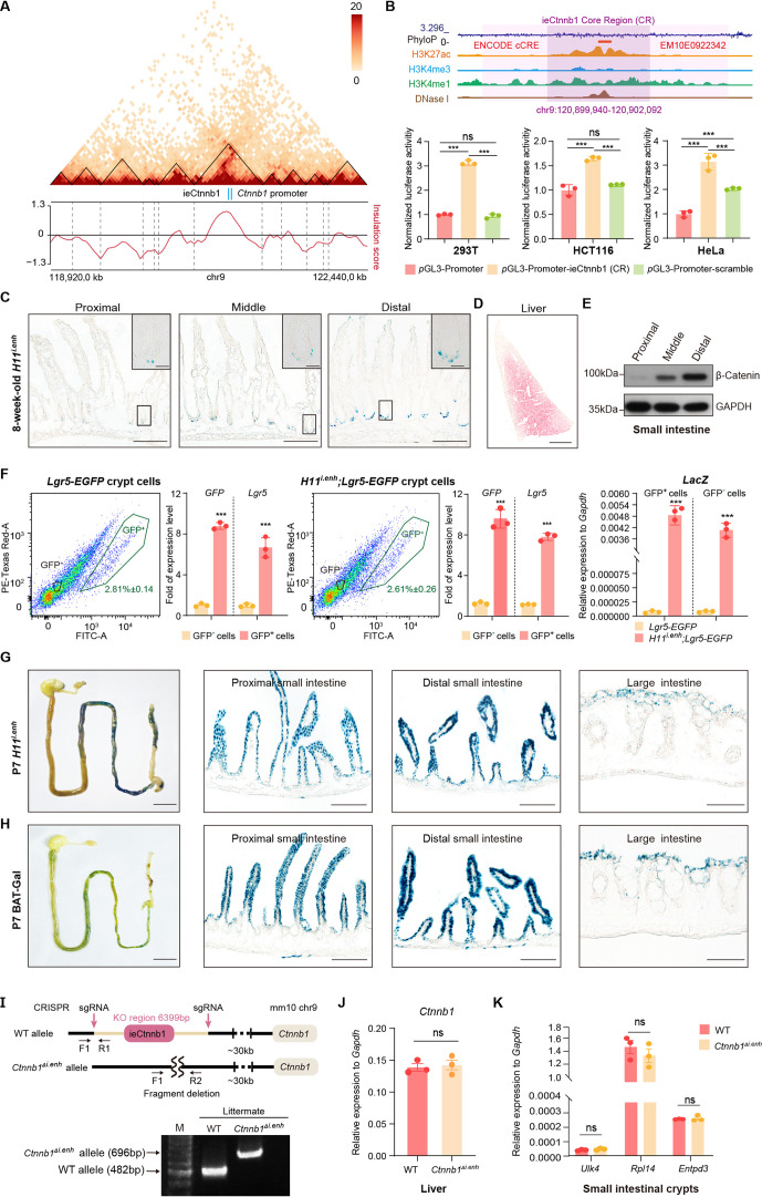
Figure 1—figure supplement 2. ieCtnnb1 is predominantly active in developing intestine.

Figure 1—figure supplement 2.

(A) Hi-C data of BALB/c mouse large intestine at indicated locus. Boundaries of topologically associating domains (TADs) and locations of Ctnnb1 promoter and ieCtnnb1 (blue bars) were marked. (B) Top: schematic representation of ieCtnnb1 core region (2,153 bp, dark purple shading). The location of an annotated ENCODE cCRE was indicated. Bottom: HEK293T, HCT116, and HeLa cells were transfected with indicated plasmids for 48 hr for luciferase reporter assay. (C) Representative images showing X-Gal staining of proximal, middle, and distal small intestinal sections of 8-week-old H11i.enh mice (blue). (D) Representative image showing X-Gal and eosin staining of the liver of 8-week-old H11i.enh mice (X-Gal: blue, eosin: red). (E) Immunoblotting of indicated proteins derived from proximal, middle, and distal small intestine tissues of WT C57/BL6 mice. (F) Flow cytometry assays to isolate GFP+ and GFP- cells from small intestinal crypts of Lgr5-EGFP (n=3) and Ctnnb1Δi.enh;Lgr5-EGFP (n=3) mice. Quantitative reverse transcription PCR (RT-qPCR) showing relative mRNA levels of GFP, Lgr5, and LacZ in aforementioned GFP+ and GFP- cells. (G–H) Representative images showing X-Gal staining of gastrointestinal tracts, and sections of proximal and distal small intestine, and large intestine of P7 H11i.enh (G) mice and P7 BAT-Gal mice (H). (I) Generation and genotyping of Ctnnb1Δi.enh mice. WT, wild-type; sgRNA, single-guide RNA. (J) Relative mRNA levels of Ctnnb1 in the liver of WT (n=3) and Ctnnb1Δi.enh (n=3) mice. (K) Relative mRNA levels of genes (Ulk4, Rpl14, and Entpd3) located in the same TAD as Ctnnb1 in small intestinal crypts of WT (n=3) and Ctnnb1Δi.enh (n=3) mice. Scale bars, 1 cm (whole mount in F and G), 100 μm (C, D, F, and G), 10 μm (magnified insets in C). Quantification data are shown as means ± SEM. Statistical significance was determined using two-way ANOVA (B) and an unpaired two-tailed Student’s t-test (F, J, and K). *p<0.05, **p<0.01, ***p<0.001, and ****p<0.0001. ns, not significant.
Figure 1—figure supplement 2—source data 1. Numerical data for Figure 1—figure supplement 2B.
Figure 1—figure supplement 2—source data 2. Numerical data for Figure 1—figure supplement 2F.
Figure 1—figure supplement 2—source data 3. Numerical data for Figure 1—figure supplement 2J.
Figure 1—figure supplement 2—source data 4. Numerical data for Figure 1—figure supplement 2K.
Figure 1—figure supplement 2—source data 5. Uncropped and labeled gels for Figure 1—figure supplement 2.
Figure 1—figure supplement 2—source data 6. Raw unedited gels for Figure 1—figure supplement 2.
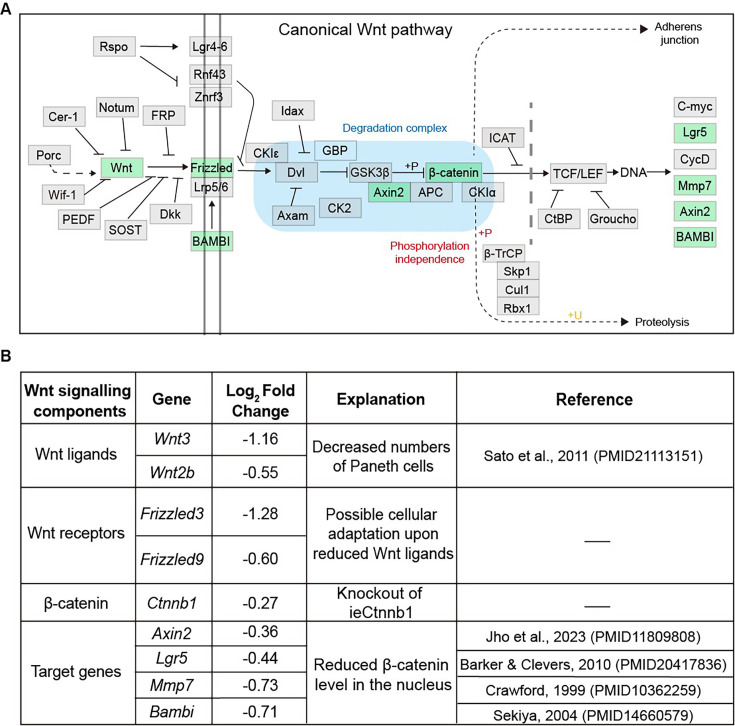
Figure 1—figure supplement 3. The list of Wnt signaling pathway components downregulated in Ctnnb1Δi.enh crypts.

Figure 1—figure supplement 3.

(A) A schematic diagram highlighting downregulated components of the Wnt signaling pathway in the crypts of the Ctnnb1Δi.enh small intestine. (B) A table showing Wnt signaling pathway components downregulated in Ctnnb1Δi.enh crypts.