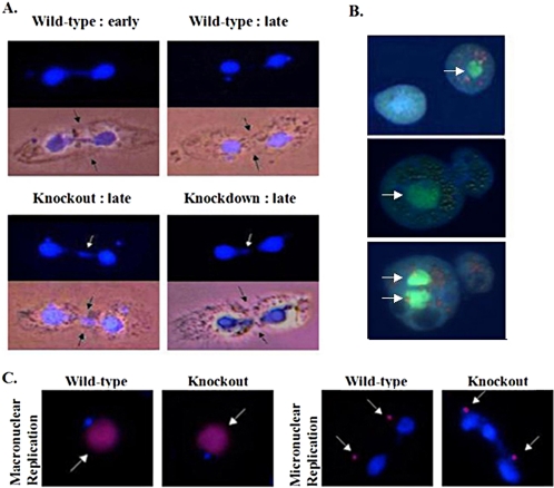
Figure 5.
Aberrant macronuclear division and cytokinesis in TIF1-deficient strains. (A) Nuclear division and cytokinesis were examined in asynchronous wild-type (CU428), homozygous TIF1 knockout (TXk202), and macronuclear TIF1 knockdown (TXh48) strains after fixation of asynchronous log phase cultures. Light images of representative predivisional wild-type cells show the typical relationship between the extent of cleavage furrow invagination (black arrows) and position of daughter macronuclei (DAPI) at early and late stages of cytokinesis. Residual macronuclear DNA at the cleave furrow in mutant cells (white arrow, late stage cytokinesis) was observed in 30–50% of mutant cell divisions. (B) Less frequent cell division phenotypes in TIF1-deficient cells. Apoflour-stained live cells were photographed immediately after cell division. Top micrograph: macronuclear division failure associated with normal cytokinesis (TXh48; arrow: macronucleus). Center micrograph: macronuclear division failure associated with asymmetric cytokinesis. Bottom micrograph: macronuclear segregation failure associated with asymmetric cytokinesis. The daughter cell on the left has two macronuclei and the one on the right has none. (C) BrdU pulse-labeling of wild-type (CU428) and homozygous TIF1 knockout (TXk202) strains. Cells were pulse-labeled for 15 min with BrdU, fixed, and stained with DAPI (blue stain) and anti-BrdU antibodies (red stain, white arrow) to examine macronuclear and micronuclear DNA replication. BrdU labeling of the macronucleus was not observed in TIF1-deficient cells undergoing aberrant macronuclear division (right micrograph).

