Figure 1.
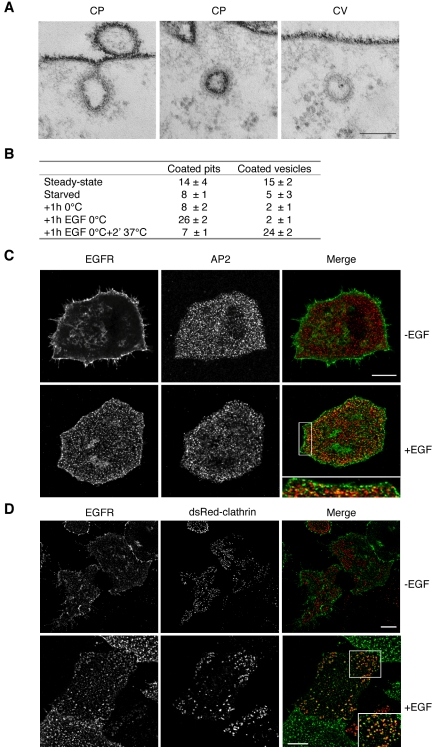
EGF stimulation induces CCPs assembly at 0°C, but not CCV budding. (A) EM images HeLa cells fixed in the presence of Ruthenium red (RR). Dark staining indicates connection with the outer surface. Examples are shown of RR-positive clathrin-coated pits (CP), and RR-negative clathrin-coated vesicles (CV). Bar, 0.06 μm. (B) Morphometric analysis of cells processed as in A. HeLa cells in logarithmic growth (“Steady state” in B) were starved in serum-free medium for 18 h at 37°C (“starved” in B), and then transferred at 0°C for 30 min, to allow the chilling of the plate and cells. The medium was then removed and cells were incubated for 1 h with fresh medium, pre-chilled on ice, in the absence (“1 h 0°C” in B), or in the presence (“1 h EGF 0°C” in B) of EGF. A parallel dish of cells, treated with EGF at 0°C as above, was then transferred for 2 min at 37°C, in the absence of EGF, to allow internalization of preformed clathrin-coated pits (“1 h EGF 0°C + 2′ 37°C” in B). Morphometry performed on 6 series of 10 cell profiles. Data represent the average number of CPs or CVs in each series, ± SD. The CP category includes: (i) all RR-positive invaginated regions of the plasma membrane, displaying a clear morphologically identifiable clathrin coat associated to the inner surface; (ii) clathrin-coated vesicle-like structures displaying a clear RR staining. (C) Double immunofluorescence showing the localization of EGFR (EGFR) and of the β subunit of the AP2 complex (AP2), in starved HeLa stimulated with EGF (+EGF) for 1 h, at 0°C, or mock-treated at the same temperature (–EGF). The right panel (merge) shows the merged images. The boxed area is displayed at higher magnification in the inset placed at the bottom of the merge panel. Bar: (bottom-right panel) 7.6 μm; (inset) 3.9 μm; (all other panels) 12.5 μm. (D) Double immunofluorescence showing the localization of EGFR (EGFR) and clathrin (dsRed-clathrin), in starved HeLa cells transfected with RFP-clathrin and stimulated with EGF (+EGF) for 1 h, at 0°C, or mock treated at the same temperature (–EGF). The right panel (merge) shows the merged images. The boxed area is displayed at higher magnification in the inset placed at the bottom of the merge panel. Bars: 12.5 μm (–EGF, all panels); 12.5 μm (+EGF); 3.9 μm (inset).

