Figure 5.
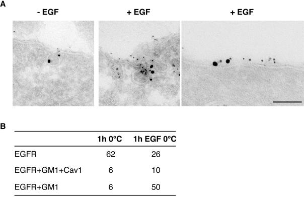
EGFR is recruited to noncaveolar, GM1-rich regions of the plasma membrane. Triple immunogold labeling of ultrathin cryosections of starved HeLa cells, labeled with CT-B/HRP, and incubated in the presence (+EGF), or in the absence of epidermal growth factor (–EGF). (A) Cryosections were triple-labeled with antibody to HRP to localize GM-1 (5 nm), anti-caveolin (10 nm), and EGFR (15 nm). The first two panels on the left show colocalization of the three antigens in caveolae. The rightmost panel shows colocalizaton of EGFR and GM1 in a flat region of the plasma membrane Bar: (left) 0.16 μm; (middle) 0.15 μm; (right) 0.1 μm. (B) Morphometric analysis of the experiment shown in A. Results show the number of 15-nm gold particles (identifying EGFR) present on the plasma membrane, colocalizing with caveolin and GM1 (EGFR+GM1+Cav1), only with GM1 (EGFR+GM1), or with neither one of the other two antigens (EGFR). Data are referred to 10 cell profiles. Of note, the triple colocalization of EGFR with GM1 and caveolin 1 was found almost exclusively within morphologically identifiable caveolae, whereas the EGFR colocalized with GM1, but not with caveolin 1, in flat regions of the plasma membrane.

