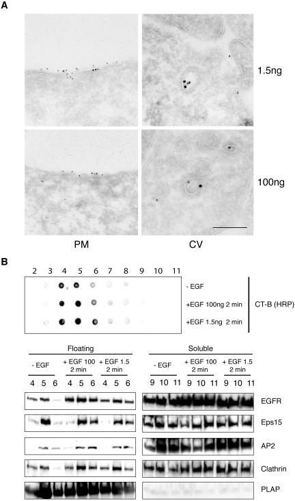
Figure 7.
EGFR recruitment and clathrin-coated pit assembly in membrane rafts are not dependent on dose and temperature of epidermal growth factor treatment. (A) Double immunogold labeling of HeLa cells treated with CT-B-HRP for 20′ at 0°C and stimulated with epidermal growth factor at 37°C for 2 min (top panels, epidermal growth factor 1.5 ng/ml; bottom panels epidermal growth factor 100 ng/ml). CT-B-HRP (10 nm) and anti-EGFR (15 nm) colocalize within discrete regions of the plasma membrane (PM), or within clathrin coated vesicles (CV). Bar: (right panels) 0.21 μm: (top left) 0.37 μm; (bottom left) 0.43 μm. (B) Starved HeLa cells were stimulated for 2 min at 37°C with epidermal growth factor at 100 ng/ml (+EGF 100), or 1.5 ng/ml (+EGF 1.5), or mock-treated (–EGF) and processed for isolation of DRMs, as described in Materials and Methods, followed by analysis by immunoblot with the indicated antibodies (bottom panel), or dot-blot (top panel) with CT-B/HRP to identify GM1. Loading of lanes was as in Figure 1.

