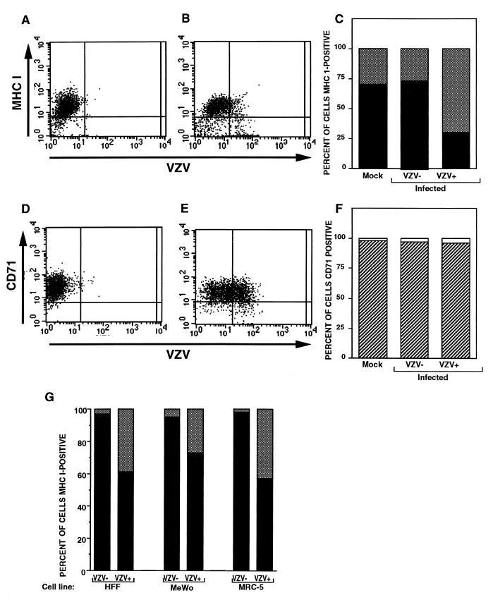
FIG. 1.
FACS analysis of MHC I molecules, transferrin receptor (CD71), and VZV proteins on VZV-infected cells. HFFs were either infected with VZV for 24 h (B and E) or mock infected (A and D), and cell preparations were stained with antibodies and fluorescent conjugates to MHC I and VZV proteins (A and B) or to transferrin receptor and VZV proteins (D and E). The percentages of VZV+ and VZV− cell populations expressing cell surface MHC I molecules (C) and transferrin receptor (F) are shown. (G) Percentage of VZV+ and VZV− cell populations expressing cell surface MHC I molecules. HFF, MeWo, and MRC-5 cells were infected with VZV for 24 h and stained with antibodies and fluorescent conjugates to MHC I and VZV proteins and analyzed by flow cytometry.

