Figure 5.
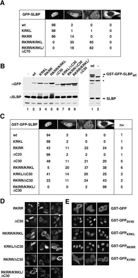
In vivo localization of SLBP mutants impaired for Impα binding. (A) Images of cells transiently transfected with the indicated GFP-SLBP construct were collected and at least 100 cells per construct were examined. Cells were scored for fusion protein localization and grouped into four categories: 1) exclusive nuclear localization; 2) greater nuclear than cytoplasmic localization; 3) even distribution between the nucleus and cytoplasm; or 4) exclusive cytoplasmic localization. (B) Western blot analysis with an anti-GFP antibody and anti-SLBP antibody, demonstrating the relative expression levels of the different GST-GFP–tagged SLBPs (top), and the levels of the endogenous SLBP as a loading control (bottom). The C-terminal GST-GFP-SLBP truncation mutants do not contain the epitope recognized by the anti-SLBP antibody necessitating their detection with the anti-GFP antibody (left, top). The expression of GST-GFP-SLBPwt (lane 1) relative to the endogenous SLBP is shown on the right. The asterisks mark the 75- and 90-kDa proteins that cross-react with the SLBP antibody. (C) Images of cells transiently transfected with the indicated GST-GFP-SLBP construct were collected and at least 300 cells per construct were examined. The number of independent transfections is indicated in the column labeled n=. Cells were scored as described in A. (D) Images of representative localization phenotypes for the indicated GST-GFP–tagged SLBP NLS mutants. (E) The predicted Imp α/β NLSs were fused to the C terminus of GST-GFP and examined for NLS activity by transient transfection. The construct transfected is indicated on the right side of each row of panels.

