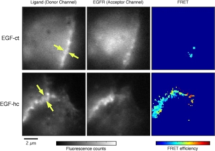
Figure 10.
FRET between ligands and the EGFR in juxtacrine complexes. Cells expressing EGF-hcF chimeras show substantial FRET signals along cell-cell contacts, indicating molecular interactions between the chimera and EGFR. Typical images of donor, acceptor and FRET efficiency, taken from cells expressing EGF-ctF (top row) and EGF-hcF (bottom row) chimeras, are shown. The right column shows images of the chimeras, tagged with labeled antibodies against the extracellular domain of EGF. The center column shows images of EGFR, tagged with the antibody against the receptor. The left column shows images of FRET efficiency, calculated pixel by pixel (see Materials and Methods).

