Figure 6.
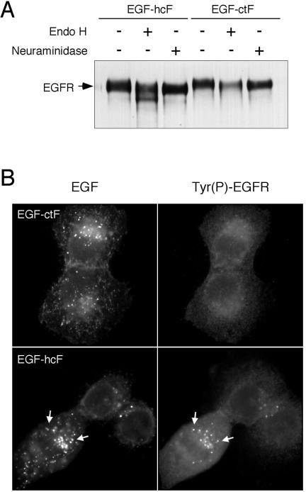
Differential glycosylation of EGFR in cells expressing EGF-ctF versus EGF-hcF. (A) The EGFR was immunoprecipitated from lysates of cells expressing either EGF-hcF (left three lanes) or EGF-ctF (right three lanes). The immunoprecipitates were treated with either endoglycosidase H or neuraminidase as described in Materials and Methods and analyzed by Western blots by using polyclonal anti-EGFR antibody SC-003. (B) Cells expressing the indicated ligand constructs were grown on coverslips, fixed, permeabilized, and stained with anti-EGF mAb (left) or affinity-purified antibodies against the major phosphorylation site in the EGFR (right) as described in Materials and Methods. Arrows indicate colocalization.

