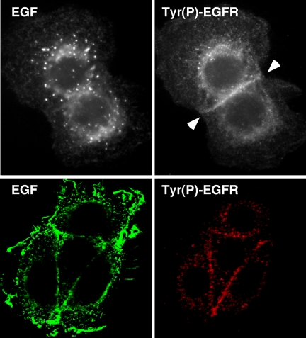
Figure 7.
Activated EGFR in cells expressing EGF-hcF are found at points of cell-cell contact. (A) Cells expressing EGF-hcF were fixed, permeabilized, and stained for both EGF (top) and tyrosine-phosphorylated EGFR as described in the legend for Figure 6B. The focal plane was adjusted to optimally visualize the area of cell-cell contact. (B) Cells were treated as described in A, but the immunofluorescence was by confocal microscopy. Shown is a group of four cells imaged through their midplane.

