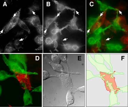
Figure 9.
Cells involved in juxtacrine signaling display extensive intercellular contacts. B82 cells expressing either the EGFR or EGF-hcF were mixed and incubated overnight in a Bioptechs coverslip dish for live cell imaging. Cells were incubated for 1 h at 37°C with 1 μg/ml anti-EGFR mAb 13A9 labeled with Alexa 488 (B, green, C and D) or anti-EGF mAb LC labeled with Alexa 594 (A, red, C and D). Arrows show areas of contact between R+ and L+ cells. Shown in E is a differential interference contrast image of D. (F) Outlined composite image of D.

