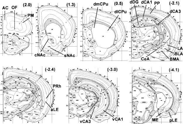
Figure 1.
Coronal mouse brain diagrams with regions of analysis indicated. Numbers in parentheses are anterior-posterior distances from Bregma in millimeters. Abbreviations of regions-for hippocampus: dCA3 and dCA1 are dorsal CA3 and CA1; vCA3 and vCA1 are ventral CA3 and CA1; for amygdala: LA, BLA, BMA, and CeA are lateral, basolateral, basomedial, and central nuclei; for striatum: dlCPu and dmCPu are dorsolateral and dorsomedial caudate/putamen; sNAc and cNAc are the shell and core regions of the nucleus accumbens; for cortex: ME, pLE, and aLE are medial, posterior lateral and anterior lateral entorhinal; PRh is perirhinal; PP is posterior parietal; OF is orbitofrontal; AC is anterior cingulate; PM is primary motor. Diagrams are reprinted with permission from Elsevier Science © 2000 (Hof et al. 2000).

