
Figure 1.
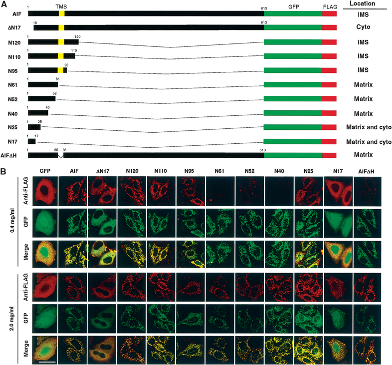
Intracellular and intramitochondrial localization of AIF variants. (A) Schematic representation of AIF variants and their intracellular or intramitochondrial localizations. The N-terminal segments of AIF from the first to the indicated residues were ligated to the N-terminus of GFP-FLAG, or the N-terminal 17 residues or TMS of AIF-GFP-FLAG was deleted (ΔN17 and AIFΔH, respectively). (B) HeLa cells expressing the indicated constructs were permeabilized and the expressed proteins were detected by GFP fluorescence and anti-FLAG antibody under confocal microscopy. Magnification, × 630; bar=20 μm.