
Figure 3.
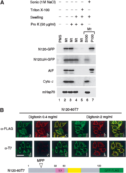
Determination of transmembrane topology of AIF. (A) The isolated mitochondria harboring N120-GFP or N120ΔH-GFP were separated into five aliquots. Four of them were incubated with or without proteinase K under the indicated conditions. The remaining aliquot was sonicated in the presence of 1 M NaCl, followed by ultracentrifugation to separate into the supernatant (S100) and membrane (P100) fractions. All these fractions were subjected to SDS–PAGE and subsequent immunoblot analysis using antibodies against the indicated proteins. (B) HeLa cells transfected with N120-GFP-FLAG carrying an internal T7-tag insertion in the residue position of 59/60 (N120-60T7) were fixed and treated with 0.4 mg/ml digitonin (a–f) or 2.0 mg/ml digitonin (g–l). The expressed proteins were detected by GFP fluorescence (green) and either anti-FLAG (shown in red; b, h) or anti-T7 antibodies (shown in red; e, k) under confocal microscopy. Merged images are also shown (c, f, i, l). Magnification, × 630; bar=20 μm.