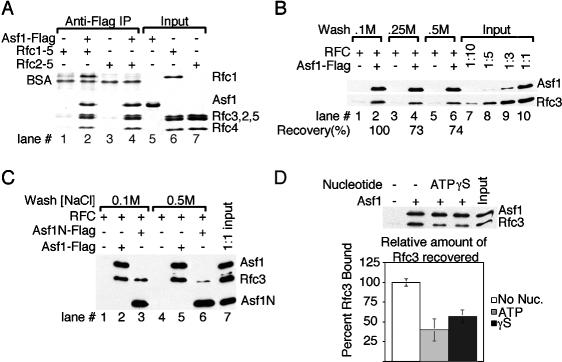
Figure 6.
RFC interacts with Asf1 in solution. (A) RFC and Rfc2-5 coprecipitate with Asf1. Fifteen picomoles of Flag-tagged Asf1 were mixed with 15 pmol of either RFC or the Rfc2-5 subcomplex prior to precipitation with anti-Flag conjugated beads. After washing with buffer + 0.1 M NaCl, precipitated polypeptides were visualized by SDS-PAGE and Coomassie staining. (B) The interaction between Asf1 and RFC is resistant to 0.5 M NaCl. One picomole of Asf1-Flag and RFC were combined in buffer with 0.1 M NaCl and precipitated as above. Precipitates were washed with buffer containing either 0.1, 0.25 or 0.5 M NaCl as indicated and recovered proteins were detected by immunoblotting. The percentage of Rfc3 recovered in each lane relative to the amount in lane 2 is presented at the bottom. (C) Both full-length and the N terminus of Asf1 interact with RFC. Immunoprecipitates were washed with either 0.1 M NaCl (lanes 1-3) or 0.5 M NaCl (lanes 4-6) and detected by immunoblotting as above. (D) Nucleotide binding partially reverses the Asf1-RFC interaction. One picomole of RFC and 1 pmol of Asf1 were combined prior to the addition of the indicated nucleotides. The amount of Rfc3 that coprecipitated with Asf1-Flag was assayed by immunoblotting. Graphed are the averages of immunoprecipitations performed in triplicate.

