
Figure 3.
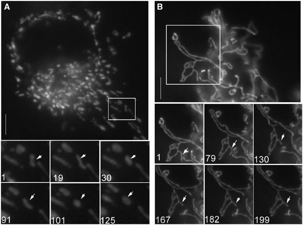
BIK induces DRP1-dependent mitochondrial fission. H1299 cells were infected for 12 h with Ad HA-BIK in presence of zVAD-fmk and in the absence (A) or presence (B) of CFP-DRP1(K38E). Mitochondria were visualized using pOCT-YFP. Images were acquired every 2 s from the boxed region of the cell presented in (A) and (B) to visualize the dynamics of mitochondrial fragments (see video 1 (panel A) and video 2 (panel B)). Bottom images represent stills taken from these movies, the numbers indicating frame number. Representative images are shown.