
Figure 2.
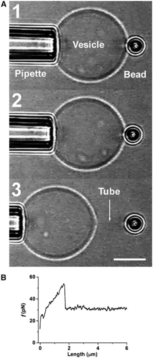
Measurement of GUV bending rigidity using a micropipette and optical tweezers. (A) (1) A GUV aspirated into a micropipette has a fixed tension. (2) The GUV containing biotinylated lipids is pressed against a 3.5 μm diameter streptavidin bead trapped by the optical tweezers. (3) The GUV is retracted and a thin tube can be formed. Bar, 10 μm. (B) A typical force–tube extension curve obtained for a 1:1:0 vesicle at a fixed tension (σ=1.3 × 10⁻⁵ N/m) during the tube extraction.