
Figure 8.
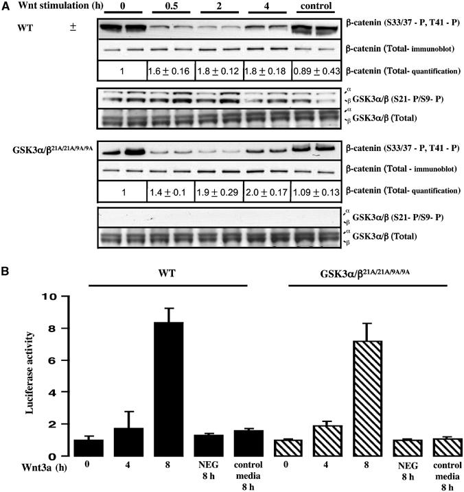
Ability of Wnt to inactivate GSK3 in knockin ES cells. (A) The WT and double knockin GSK3α/β21A/21A/9A/9A ES cells were incubated in medium containing Wnt3a or a control medium obtained from non-Wnt3a-expressing cells. At the indicated times, the phosphorylation state of β-catenin was assessed by immunoblot analysis employing an antibody that recognises β-catenin when phosphorylated on the three residues targeted by GSK3 (S33/37-P, T41-P). Quantitation of the total levels of β-catenin was performed by immunoblot analysis using LI-COR Odyssey infrared imaging system. The extracts were also immunoblotted with antibodies recognising the phosphorylated and total forms of GSK3. For each condition duplicate dishes were analysed and similar results were obtained in two separate experiments. (B) ES cells were co-transfected with a firefly luciferase reporter construct containing LEF/TCF-binding sites as well as a constitutive SV-40 Renilla luciferase construct, to control for transfection efficiency. At 24 h post-transfection, cells were incubated in medium containing Wnt3a or a control medium obtained from non-Wnt3a-expressing cells and luciferase activity was measured. As a control, cells were transfected with a firefly luciferase reportor construct in which the LEF/TCF-binding sites had been mutated (NEG) and were incubated with Wnt3a for 8 h. Luciferase activity was expressed in arbitrary units relative to the activity observed in unstimulated WT cells normalised for Renilla luciferase activity. Four dishes of cells were analysed for each condition and similar results were obtained in two separate experiments. Results shown are mean±s.d. for a single experiment.