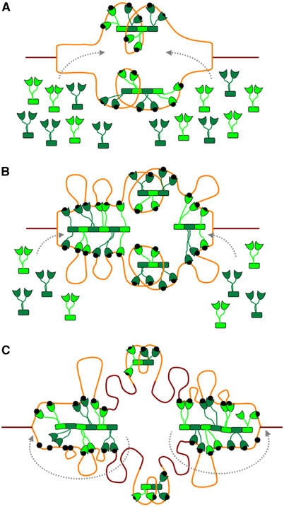
Figure 6.

Model of SeqA sequestration and SeqA foci migration following the replication forks. Fully and hemimethylated DNA are shown in brown and orange, respectively. Black dots represent hemimethylated GATC sequences. SeqA dimers are shown in green. (A) As replication initiates, SeqA binds newly generated hemimethylated GATC sequences within oriC, triggering origin sequestration. (B) As the forks progress, more hemimethylated GATC sequences become available. Spacing between the newly occurring GATC sequences favors SeqA filament formation, which in turn favors cooperative binding of distant GATC sequences. (C) As cells have a limited amount of SeqA, dynamic addition and removal of SeqA dimers (indicated by dashed gray arrows) will allow the SeqA filament to bind newly occurring hemimethylated GATC sequences and track forks as they progress. Only few SeqA molecules are required for origin sequestration and, therefore, oriC will remain hemimethylated while the intervening DNA sequences are remethylated.
