
Figure 3.
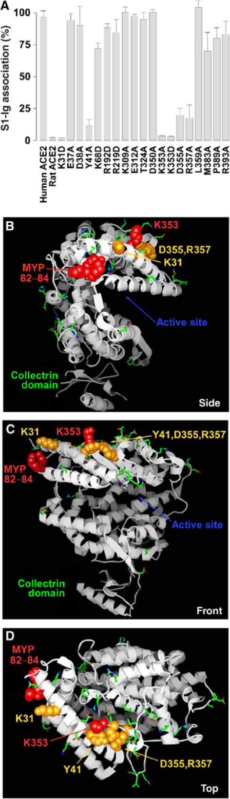
The S-protein-binding site of human ACE2. (A) Experiments identical to those of Figure 1 except that the indicated solvent-accessible residues common to human and rat ACE2 were modified in human ACE2 to either alanine or aspartic acid. (B) Representation of the crystal structure of human ACE2, with the collectrin domain oriented downward and viewed from the side of the cleft bearing the enzymatic active site. Residues of rat ACE2 whose alteration to the corresponding human residues converted rat ACE2 to an efficient SARS-CoV receptor are shown in red. Human ACE2 residues whose alteration substantially decreased S1-Ig association are shown in orange. Residues whose alteration did not affect S1-Ig association are shown in green. Low-resolution electron density associated with the collectrin domain is represented by a small β-sheet and α-helix at the base of the figure. (C) A view identical to that in (B) except that the molecule has been rotated 90° about the vertical axis. (D) A view identical to that in (C) except that the molecule has been rotated 90° about the horizontal axis.