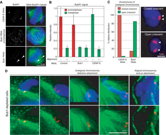
Figure 5.

Bub1 depletion leads to defective MT–kinetochore attachments. (A) Immunofluorescence images of metaphase CENP-E-depleted (upper panel), prometaphase Bub1-depleted (middle panel) and metaphase Bub1-depleted (bottom panel) cells stained with DAPI (blue), anti-BubR1 antibodies (red) and tubulin antibodies (green). To arrest cells at metaphase, they were treated with MG132. Scale bar: 10 μm. Images of cells stained in parallel were acquired at the same time to assist comparisons across samples. Note that BubR1 levels on kinetochores in Bub1-depleted metaphase cells are low, implying that the kinetochores are MT-bound; high BubR1 levels in prometaphase demonstrate that BubR1 can bind kinetochores of unattached chromatids even when Bub1 is depleted. (B) Quantification of BubR1 levels on individual kinetochores from the data in (A). Error bars indicate s.e.m. Prometaphase and metaphase data are necessarily derived from different cells, but unaligned and aligned data for Bub1 and CENP-E depletions in metaphase were derived from the same cells. (C) Quantification of immunofluorescence images of unaligned chromosomes in Bub1- and CENP-E-depleted cells arrested for 1 h at the metaphase–anaphase transition with MG132, stained for DNA (blue) and BubR1 (red). Kinetochores were classified as having either a closed (upper panel) or open crescent morphology (lower panel). Scale bar: 1 μm. (D) Immunofluorescence images of unaligned chromosomes in Bub1-depleted cells arrested at the metaphase–anaphase transition for 1 h with MG132, stained with DAPI (blue), anti-CENP-E antibodies (red) and anti-β-tubulin antibodies (green). Note the MTs running past the open crescent-shaped kinetochores. As a comparison, end-on attached kinetochores of correctly aligned sister chromatids are shown. Scale bar: 5 μm.
