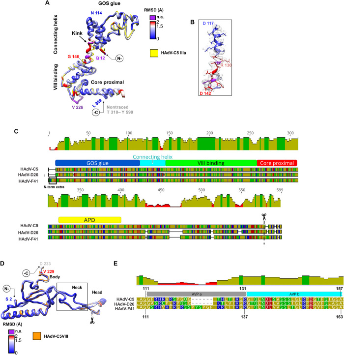
Fig. 5. Proteins IIIa and VIII.
(A) Superposition of HAdV-C5 IIIa (PDB ID: 6B1T, chain N) in yellow and HAdV-F41 IIIa colored by RMSD as in Fig. 2. The GOS glue, connecting helix, VIII-binding, and core proximal domains are indicated. (B) Detail of the connecting helix to show its fit into the cryo-EM map. (C) Schematics showing the alignment of protein IIIa sequences in HAdV-C5, HAdV-D26, and HAdV-F41. Traced domains are indicated below, in different colors. APD is the appendage domain traced in HAdV-D26, but not in the other two viruses. The N-terminal extension in HAdV-F41 (N-term extra) and maturation cleavage site (scissors) are also indicated. (D) Superposition of HAdV-C5 protein VIII (PDB ID: 6B1T Chain O) in orange and HAdV-F41 VIII colored by RMSD. The body, neck, and head domains are indicated, as well as the gap corresponding to the peptide cleaved during maturation (scissors). (E) Sequence alignment showing the two central peptides of protein VIII cleaved by AVP (AVPa and AVPb) in HAdV-C5, HAdV-D26, and HAdV-F41. Amino acid and mean pairwise identity histogram color schemes are the same as in Fig. 2.

